CERVICAL PAIN: EXERCISES AND STRETCHES

pain


Cervical pain is one of the most common injuries in people of all ages, being one of the most prevalent conditions in Western society. This problem can be derived from a seating during long periods of time, aggravated by a greater tendency to the use of smartphones, of the computer, use of chairs and tables not suitable for each person and a sedentary lifestyle.

This medical condition can be caused by maintaining an forward head posture (FHP), it is characterized by an excessively advanced position of the head with respect to the neck and an internal rotation of the shoulders.

FHP is associated with low cervical flexion (C4-C7) and high cervical hyperextension (C1-C3), generating musculoskeletal changes at the cervical level. The deep neck muscles are considered very important in the stability, support and adjustment of the neck posture. Generally, there is a weakness of the deep flexor neck muscles in addition to a lack of strength of the external retractors and rotators of the shoulder. To solve this weakness, the body must generate compensations producing a shortening of the high fibers of the trapezium, sternocleidomastoid, scapula elevator, pectoralis major and minor and extensor musculature of the head.

Bad cervical posture can lead to a limitation of mobility and cause excessive tension of muscles and soft tissues. People with neck pain tend to move their head forward with respect to the neck without realizing it. In addition, previous studies have associated FHP and shoulders in internal rotation with cervical and headaches.

To correct this posture, it has been observed that proper activation of the deep flexor muscles of the neck during craniocervical flexion helps maintain an upright posture of the head. Therefore, the strengthening of weakened muscles and stretching of the trapezius, sternocleidomastoid and scapula lift can have positive effects on FHP and cervical pain.

Given the aforementioned consequences of the FHP, it seems correct to create and disseminate an exercise program to correct these dysfunctions.

Video on YouTube with explanation about the exercises and stretching that we explain below:


Strength exercises


Perform 3 sets of 10 repetitions in each exercise, the shoulders and neck should be at the beginning in a relaxed position. That is, open chest, shoulders away from the ears and slight cervical flexion


1. Cervical flexion


Face up with the head resting on the floor, perform a cervical flexion reducing the distance between the chin and the sternum. We activate and train the deep neck muscles. (Image 1)

Image 1: Deep neck flexors exercise


2. External rotation


In lateral recumbency with the elbow resting on the side, perform an external shoulder rotation movement making a movement towards the side of the hand that moves away from the body. Use a hand-held weight to get more training in the rotator muscles of the shoulder. (Image 2)


Image 2: External shoulder rotators exercise


3. T shape


When standing with legs bent, trunk tilted forward, back straight and arms stretched to the ground, abduct both arms with elbows extended to a position of 90潞 with respect to the body forming a T. Use weights on both arms to improve strength in the external abductor and rotator musculature of the shoulder, in addition to scapula approximators and stabilizers (Image 3 and 4)

Image 3: Abduction shoulder exercise with extended elbows (front view)


Image 4: Abduction shoulder exercise with extended elbows (sagittal view)


4. W shape


Standing with your legs bent, trunk tilted forward, back straight, shoulders adducted on your chest, elbows bent 100 ° and palms up, abduct your shoulders to back height forming a W. Use weights on both arms to increase training in the external abductor, flexor and rotator muscles of the shoulder, as well as scapula approximators and stabilizers. (Image 5 and 6)


Image 5: Abducted shoulder elbow flexion exercise (front view)


Image 6: Abduction shoulder exercise flexed elbows (sagittal view)


Stretching


Perform each stretch 30 seconds.


1. Pectoral


With the forearm resting on a wall and the shoulder at 90 °, make a rotation with the body in the opposite direction to the arm so that we achieve a separation between the origin and insertion of the pectoral muscle. (Image 7)

Image 7: Pectoral stretching (anterior and posterior view)


2. Upper trapezius


Perform a cervical flexion (look down), contralateral tilt (bring the ear to the shoulder) and homolateral rotation, with the contralateral hand increase the position of the neck to help increase tension in the trapezius. (Image 8)


Image 8: Trapeze Stretch


3. Neck Extenders


Perform a pure cervical flexion (look down), with both hands passively increase the position of the neck until tension is felt in the posterior area of ​​the neck. (Image 9)

Image 9: Stretching the neck extenders


DOLOR CERVICAL: EJERCICIOS Y ESTIRAMIENTOS


dolor


El dolor cervical es una de las lesiones m谩s comunes en personas de todas las edades, siendo una de las condiciones m谩s prevalentes en la sociedad occidental. Este problema puede ser derivado de una sedestaci贸n durante periodos largos de tiempo, agravado por una mayor tendencia al uso de smartphones, del ordenador, uso de sillas y mesas no aptas a cada persona y un estilo de vida sedentario.

Esta dolencia puede ser provoca por mantener una postura de la cabeza adelantada (PCA), se caracteriza por una posici贸n excesivamente adelantada de la cabeza respecto al cuello y una rotaci贸n interna de los hombros.

La PCA est谩 asociada a una flexi贸n cervical baja (C4-C7) y una hiperextensi贸n cervical alta (C1-C3) generando cambios musculoesquel茅ticos a nivel cervical. La musculatura profunda del cuello es considerada muy importante en la estabilidad, soporte y el ajuste de la postura del cuello. Generalmente, se produce una debilidad de la musculatura flexora profunda del cuello adem谩s de una falta de fuerza de los retractores y rotadores externos del hombro. Para solventar esta debilidad, el cuerpo debe generar compensaciones produciendo un acortamiento de las fibras altas del trapecio, esternocleidomastoideo, elevador de la esc谩pula, pectoral mayor y menor y musculatura extensora de la cabeza.

Una mala postura cervical puede derivar en una limitaci贸n de movilidad y provocar una tensi贸n excesiva de m煤sculos y tejidos blandos. Las personas con dolor de cuello tienden a desplazar su cabeza hacia delante con respecto al cuello sin darse cuenta. Adem谩s, estudios previos han asociado la PCA y hombros en rotaci贸n interna a dolores cervicales y de cabeza.

Para corregir esta postura, se ha observado que una correcta activaci贸n de los m煤sculos flexores profundos del cuello durante la flexi贸n craneocervical ayudan a mantener una postura erguida de la cabeza. Por lo que, el fortalecimiento de la musculatura debilitada y el estiramiento del trapecio, esternocleidomastoideo y elevador de la esc谩pula puede tener efectos positivos sobre la PCA y el dolor cervical.

Teniendo en cuenta las consecuencias antes mencionadas de la PCA, parece correcto crear y difundir un programa de ejercicios para corregir estas disfunciones.

V铆deo en Youtube con explicaci贸n sobre los ejercicios y estiramientos que a continuaci贸n explicamos:


Ejercicios de fuerza


Realizar en cada ejercicio 3 series de 10 repeticiones, los hombros y cuello deben de estar al inicio en una posici贸n relajada. Es decir, pecho abierto, hombros lejos de las orejas y una ligera flexi贸n cervical


1. Flexi贸n cervical


Boca arriba con la cabeza apoyada en el suelo, realizar una flexi贸n cervical reduciendo la distancia entre la barbilla y el estern贸n. Activamos y entrenamos la musculatura profunda del cuello. (Imagen 1)
 
Imagen 1: Ejercicio flexores profundos del cuello


2. Rotaci贸n externa


En dec煤bito lateral con el codo apoyado en el costado, realizar un movimiento de rotaci贸n externa de hombro realizando un movimiento hacia el lateral de la mano que se aleja del cuerpo. Utilizar una pesa cogida con la mano para conseguir un mayor entrenamiento en los m煤sculos rotadores del hombro. (Imagen 2)


Imagen 2: Ejercicio rotadores externos del hombro


3. Forma de T


En bipedestaci贸n con las piernas flexionadas, tronco inclinado hacia delante, espalda recta y brazos estirados hacia el suelo, abducir ambos brazos con los codos extendidos hacia una posici贸n de 90潞 con respecto al cuerpo formando una T. Utilizar pesas en ambos brazos para mejorar la fuerza en la musculatura abductora y rotadora externa del hombro, adem谩s de aproximadores y estabilizadores de la esc谩pula (Imagen 3 y 4)

 
Imagen 3: Ejercicio abducci贸n del hombro con codos extendidos (vista frontal)


Imagen 4: Ejercicio abducci贸n del hombro con codos extendidos (vista sagital)


4. Forma de W


De pie con las piernas flexionadas, tronco inclinado hacia delante, espalda recta, hombros aducidos sobre el pecho, codos flexionados 100° y palmas de las manos hacia arriba, abducir los hombros hasta la altura de la espalda formando una W. Utilizar pesas en ambos brazos para aumentar el entrenamiento en la musculatura abductora, flexora y rotadora externa del hombro, adem谩s de aproximadores y estabilizadores de la esc谩pula. (Imagen 5 y 6)


Imagen 5: Ejercicio abducci贸n del hombro codos flexionados (vista frontal)


Imagen 6: Ejercicio abducci贸n del hombro codos flexionados (vista sagital)


Estiramientos


Realizar cada estiramiento 30 segundos.


1. Pectoral


Con el antebrazo apoyado sobre una pared y el hombro a 90°, realizar una rotaci贸n con el cuerpo en direcci贸n opuesta al brazo para que logremos una separaci贸n entre el origen e inserci贸n del m煤sculo pectoral. (Imagen 7)


Imagen 7: Estiramiento del pectoral (vista anterior y posterior)


2. Trapecio superior


Realizar una flexi贸n cervical (mirar hacia abajo), inclinaci贸n contralateral (llevar la oreja hacia el hombro) y rotaci贸n homolateral, con la mano contralateral aumentar la posici贸n del cuello para ayudar a aumentar la tensi贸n en el trapecio. (Imagen 8)


Imagen 8: Estiramiento del trapecio


3. Extensores de cuello


Realizar una flexi贸n cervical pura (mirar hacia abajo), con ambos manos aumentar pasivamente la posici贸n del cuello hasta sentir tensi贸n en la zona posterior del cuello. (Imagen 9)


Imagen 9: Estiramiento de los extensores del cuello


RUNNING: MAIN INJURIES


In recent years, running has increased its popularity in our society, breaking records of participation in various popular races. In each of our cities we can observe a large number of “runners” alone or in groups trying to improve their brands or simply enjoying the sport.
Most runners have suffered an injury throughout their lives, covering problems from the foot to the cervical level. In this article we will analyze the most common injuries in runners and how to prevent or treat them.

The body gives us warning signs, the following phrases will sound: "Uff, how loaded I am", "today it was difficult for me to get in bed", "I have noticed a pull on the leg"
Physiotherapy can help us not to say these phrases or, if it is too late, to prevent further injury. Listen to your body and consult your physiotherapist with any questions and do not hesitate to call us for any questions.

The most common injuries are: plantar fasciitis, iliotibial girdle, tibial periostitis, tedinopathies, sprains and muscle tears.

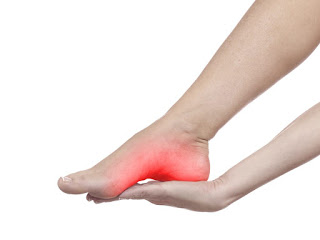
- Plantar fasciitis: the plantar fascia is defined as a fibrous and elastic tissue, which covers most of the sole of the foot, and that originates in the calcaneus bone (heel bone), until it is inserted into the base of the metatarsals (fingers) It is responsible for stabilizing and supporting the entire foot assembly; joints and plantar arch.

Plantar_fasciitis

    There are several causes or risk factors for which it can occur are: the increase in physical or sports load, or sudden change in it; plantar arch alteration, whether increased or decreased (cavus or flatfoot); tension or retraction of the Achilles tendon; inappropriate footwear; Obesity or sudden weight gain.


- Iliotibial waist: it is an inflammation that occurs in the lateral or external area of ​​the knee, caused by the friction caused between the lateral condyle of the femur and the iliotibial waistband. It is usually a very typical pain in runners, due to bad biomechanics of the race when the muscles are fatigued. The main problem is the failure of the gluteus medius in monopodial support causing the knee to go further in each step and the friction described above occurs.


- Shin Splints: pain located along the two distal thirds of the posterior medial tibia during exercise, with pain in the palpation of the tibia. It is caused by overloading of the tibial bone cortex, resulting in an inflammation of the membrane that covers the bone.


       The risk factors by which it can occur are: increase in body mass; non-progressive increase in training; flat foot, a bad biomechanics of tread produces adaptations of other areas of the body and can affect the tibia; lack of range of motion in the ankle; lack of range of movement in the hip: the lack of strength in the gluteus medius triggers a valgus knee and, as a consequence, an incorrect tread; run on hard surfaces.


- Tendinopathies: Tendonitis is a very frequent pathology, which usually appears after excessive efforts, repetitive movements and direct trauma. It consists of the inflammation of the tendons, which are resistant strings of tissue that link the muscles with the bones and help the muscles move the bones.

Avoid repetitive movements and overload, keep the muscles strong and flexible, exercise the upper and lower extremities, without excessively repeating the same movement, alternating different types of movements, with small recovery intervals. Good hydration is also recommended, as it always helps to maintain a better vascularization of the area.


- Sprains: it is an elongation or rupture of the ligaments that slow the inversion of the foot. We must perform balance or proprioception exercises to improve the position of the ankle at the nervous system level and strengthen the muscles that are responsible for maintaining a correct positioning of the foot.


- Muscle tears: it is the separation or lack of congruence of muscle fibers. Whether you are a sports person or if you are more sedentary, a key aspect is to keep the body hydrated. If any sporting activity is carried out, it is essential to perform a good warm-up prior to it, because that way we can prepare the muscles for the activity, so that they can react quickly and correctly.
In addition, resting times are essential. If we do not rest properly between workouts we will have fatigued muscles, so that they will be unable to adapt effectively to the activity to which we are subjected. It is very important to let the muscle recover. In the case of runners, muscle overload can lead to muscle breakage due to changes in pace or high running intensities. That is why it is very important to treat the affected muscles to prevent it from going further. The body is warning us and we must listen.


KNEE OSTEOARTHRITIS: EXERCISES


knee

WHAT IS IT?

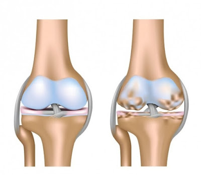
Osteoarthritis, or wear of the knee joint, is a painful, non-inflammatory, irreversible degenerative disease of the knee joint caused by wear of the articular cartilage.
Osteoarthritis of the lower limb is a common condition that affects the elderly, approximately 20% of people over 60 years of age worldwide experience knee pain. It represents 80% - 90% of hip and knee replacements in the United States and the United Kingdom.


KNEE ANATOMY

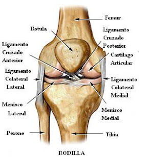
The knee is formed by the union of 2 important bones, the femur in its distal portion, and the tibia in the proximal portion. It also has a small bone, called the patella, which articulates with the anterior and inferior portion of the femur. It can mainly perform flexion and extension movements. It is surrounded by an articular capsule and several ligaments that give it stability. In its vicinity, powerful muscles are inserted that make limb movement possible.
It is composed of the joint action of the femur, tibia, patella and two fibrocartilaginous discs that are the meniscus. Femur and tibia make up the main body of the joint, while the patella acts as a pulley and serves as an insertion to the quadriceps muscle tendon and the patelar tendon whose function is to transmit the force generated when the quadriceps is contracted.

anatomy_knee


PROTHESIS

A knee prosthesis is a mechanical element composed of various metal and plastic components that replace the knee joint consisting of tibia, femur and kneecap.
Should I wear a prosthesis? No, we must understand that osteoarthritis is a degeneration of the joint due to its use. Over the years, all people have osteoarthritis to a greater or lesser extent. Osteoarthritis is NOT an indication of putting on a prosthesis, the indication of putting on the prosthesis is given by pain and / or the functional limitation produced by this osteoarthritis.

prothesis_knee


CAUSES

The main causes of osteoarthritis of the knee are:
Trauma during sports and recreational activities, hard physical work overload, congenital joint deformity, metabolic disorders, overweight.


TREATMENT


The guideline for the treatment of osteoarthritis is exercise as a non-pharmacological therapy. Exercise improves the symptoms and general well-being of people with this pathology, while they are relatively safe as compared to pharmacological treatments. The improvement of pain and functional results after treatment with osteoarthritis exercises are demonstrated by numerous studies.
Great benefits, regarding pain and function, were observed in people with osteoarthritis of the knee who exercised. The effectiveness of the exercise was generally greater at 2 months after starting the exercise, according to the results of the studies.
For the treatment of osteoarthritis pain we also use radiofrequency, deep heat that helps relieve nerve endings and relax symptoms.


MAIN FACTORS


- The age of who suffers from this deficiency affects, trials with younger participants demonstrated a more appreciable functional improvement than older patients after exercise treatment. In the older population, other age-related conditions (eg, reduced functions in the cardiovascular and musculoskeletal systems) may also explain the observed effect.

- Osteoarthritis severity: in general, the results support the inverse association between benefits of exercise and the severity of osteoarthritis of the knee (that is, it is believed that the exercise produces a greater improvement with a milder knee osteoarthritis than more severe). Patients on the waiting list for surgery, who have knee osteoarthritis in the most advanced stage of the clinical spectrum, showed a smaller exercise response compared to those who were not on a waiting list.


EXERCISE PROGRAM


We will perform the exercises 3 sets of 10 repetitions each, 3 times / week.

Exercise 1:

Lying on your back with your leg straight and a towel under your knee, perform quadriceps contractions by crushing the towel with a hollow of your knee. Bring the toe towards us to feel more muscle contraction.


Exercise 2:

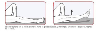
Lying on your back with your leg straight, raise it straight until you reach about 45 degrees from the ground, bringing the tip of the foot up and down again, relaxing the tip of the foot.


Exercise 3:

Lying on your side with your hips and knees bent, separate your knees from each other but not your feet Being able to increase the resistance by placing an elastic band between the knees. We want to focus on strengthening the buttocks.


Exercise 4:

Sitting on a high surface, make knee extensions bringing the tip of the foot towards us at the end of the movement and withstand that tension for 5 seconds.


Exercise 5:

Standing with one leg elevated and the support leg slightly bent, balance for 20-30 seconds and rest for a few seconds. If it seems easy you can increase the difficulty by making circles with the leg that is in the air, closing your eyes, using unstable surfaces ...


SCIATICA RELATED TO HERNIATED DISC: SURGERY OR CONSERVATIVE TREATMENT


herniated-disc-sciatic

Herniated disc and sciatic


The column vertebrae are formed with 7 cervical vertebrae, 12 thoracic, 5 lumbars, 5 sacral and 3 coccygeus. Between each vertebra there is a disc formed with the annulus fibrosus and nucleus pulposus giving movement to the back, support and cushion the body´s weight.


The lower back is more prevalent to have a herniated disc having to support more weight than the other vertebrae and because it has a high mobility in flexion. The area of least resistance offered by the fibrous annulus is the posterior part. The nucleus is slightly delayed with respect to the geometric centre of the disc. This situation together with the bending movement causes the nucleus to exert more pressure on the back of the annulus.


Over the years, annulus wear down and nucleus can to go out. Usually, the herniated disc is not symptomatology but if nucleus affects the sciatic nerve we can feel symptoms. The most important symptom of sciatica is lumbosacral radicular leg pain that follows a dermatomal pattern radiating below the knee and into the foot and toes (1, 2).


lumbar-disc
Lumbar vertebrae


(Information extracted from: Jacobs, Wilco CH, et al. "Surgery versus conservative management of sciatica due to a lumbar herniated disc: a systematic review." European Spine Journal 20.4 (2011): 513-522)

The prevalence of sciatic symptoms reported in the literature varies considerably ranging from 1.6% in the general population to 43% in a selected working population (3). Although the prognosis is good in most patients (4), a substantial proportion (up to 30%) continues to have pain for 1 year or longer (5, 6).


 The goal of this study was to assess the effects of surgery versus conservative therapy (including epidural injections) for patients with sciatica due to lumbar disc herniation.


 In total, five studies were identified. One study compared early surgery with prolonged conservative care followed by surgery if needed (7); three studies compared surgery with usual conservative care (8,9,10), and one study compared surgery with epidural injections (11).
  

Early surgery versus prolonged conservative care


 1.       Relief of leg pain was faster for patients assigned to early surgery. Intention to treat analysis
showed statistically significant more leg pain relief in favour of early surgery as compared with prolonged conservative care at 3 months. There was no significant overall difference between the two groups in disability scores during the first year. The median time to recovery was 4 weeks for early surgery and 12 weeks for prolonged conservative treatment (7).


Surgery versus usual conservative care

 2.       Both patient and observer ratings demonstrated that discectomy was significantly better than conservative treatment at 1 year. After 1 year, 24 of the 66 patients (36%) in the conservative care group versus 39 of the 60 patients (65%) in the surgery group reported a good outcome. No significant differences in outcomes were reported at 4 and 10 years follow-up (9).


 3.       Overall, no significant differences were found for leg pain or back pain, and subjective disability throughout the 2 years of follow up. Visual analog scale leg pain scores, however, improved more rapidly in the discectomy group first 6 weeks (8).


 4.       Confirmed disc herniation showed that both the surgery as well as the conservative treatment group improved substantially over 2 years of leg pain and/or back pain (10).
  

Surgery versus epidural steroid injections


 5.       Patients undergoing discectomy had the most rapid decrease in their symptoms. The decrease in leg pain in the discectomy group was significantly greater than in the epidural steroid injection group at 3 and 6 month follow-up intervals, but not beyond 1 year. There were no significant differences between groups for back pain throughout the follow-up (11).


sciatic-steroid
Epidural steroid injection


Conclusions


 In general, there is evidence that early surgery in patients with sciatica provides for a better short-term relief of leg pain as compared to prolonged conservative care. But no significant differences were found between surgery and usual conservative care in any of the clinical outcomes after 1 and 2 years.


Bibliography


 1. Valat JP, Genevay S, Marty M, Rozenberg S, Koes B (2010) Sciatica. Best Pract Res Clin Rheumatol 24:241–252

 2. van Tulder M, Peul W, Koes B (2010) Sciatica: what the rheumatologist needs to know. Nat Rev Rheumatol 6:139–145


 3. Konstantinou K, Dunn KM (2008) Sciatica: review of epidemiological studies and prevalence estimates. Spine (Phila Pa 1976) 33:2464–2472


 4. Legrand E, Bouvard B, Audran M, Fournier D, Valat JP (2007) Sciatica from disk herniation: medical treatment or surgery? Joint Bone Spine 74:530–535


 5. Vroomen PC, de Krom MC, Slofstra PD, Knottnerus JA (2000) Conservative treatment of sciatica: a systematic review. J Spinal Disord 13:463–469


 6. Weber H, Holme I, Amlie E (1993) The natural course of acute sciatica with nerve root symptoms in a double-blind placebocontrolled trial evaluating the effect of piroxicam. Spine (Phila Pa 1976) 18:1433–1438


 7. Peul WC, van Houwelingen HC, van den Hout WB, Brand R, Eekhof JA, Tans JT, Thomeer RT, Koes BW (2007) Surgery 
versus prolonged conservative treatment for sciatica. N Engl J Med 356:2245–2256

 8. Osterman H, Seitsalo S, Karppinen J, Malmivaara A (2006) Effectiveness of microdiscectomy for lumbar disc herniation: amrandomized controlled trial with 2 years of follow-up. Spine (Phila Pa 1976) 31:2409–2414

 9. Weber H (1983) Lumbar disc herniation. A controlled, prospective study with ten years of observation. Spine (Phila Pa 1976) 8:131–140


 10. Weinstein JN, Tosteson TD, Lurie JD, Tosteson AN, Hanscom B, Skinner JS, Abdu WA, Hilibrand AS, Boden SD, Deyo RA 
(2006) Surgical vs nonoperative treatment for lumbar disk herniation: the Spine Patient Outcomes Research Trial (SPORT): a randomized trial. JAMA 296:2441–2450

 11. Buttermann GR (2004) Treatment of lumbar disc herniation: epidural steroid injection compared with discectomy. A prospective, randomized study. J Bone Joint Surg Am 86-A:670–679

ANKLE SPRAIN: TREATMENT


Sprained-ankle

Ankle sprain is one of the most common injuries in contact sports or that involve jumps like basketball or soccer. The ligament injuries happen generally for an acute traumastism, rarely occur for overuse. An ankle sprain occurs when the strong ligaments that support the ankle stretch beyond their limits and tear when we do an inversion of our foot. 


Anatomy


The ankle has 3 ligaments in its external part: anterior talofibular, calcaneofibular, posterior talofibular. All of them can be injuried, but the common ligament affected is the first one in a 70% of the time because it is the most exposed to the stretch. 

 
ligaments-ankle
Ligaments of the ankle

Classification of ligaments injuries


Grade I: Mild. It represents a microscopic injury without stretching of the ligament on a macroscopic level.

Grade II: Moderate. It has macroscopic stretching, but the ligament remains intact.

Grade III: Severe. It is a complete rupture of the ligament. 


Treatment


When a ligament is stretched or broke occurs a vessels constriction around 10-15 minutes followed by an inflammation caused by increased blood flow to the area in the next 48-72 hours. Furthermore due the immobilisation, our muscles lost strength and the nervous system miss proprioception though which we perceive the position and movement of our body, including our sense of equilibrium and balance. The next treatment will fix these symptoms and problems. 


Acute phase (first 48-72 hours)


R: Rest. Avoid any activity that may aggravate the injury.

I: Ice. Maintain the application of cold 20 minutes being able to repeat the operation 3 and 5 times a day allowing at least 2 hours between each one.

C: Compression. Elastic tape for to protect the injury.

E: Elevation. Purpose is to avoid inflammation. 

 
RICE


Range of movement (since 4潞-7潞 day)


Two series of 10 repetitions 2 times per day.


Active: lie on your back do flexion dorsal, flexion plantar, inversion, eversion and alphabetic movements (1 minute). 


plantar-flexion-dorsal-inversion-eversion
Active movements without weight


Isometric: sit down in a chair do flexion plantar with the other foot on, Inversion, eversion against a wall and thrusts towards the ground. 


isometric
Isometric movements without weight


Strength (since 15潞 day)


Two series of 10 repetitions 2 times per day.


Isometric: stand up do flexion plantar, Inversion, eversion and thrusts towards the ground


Active: lie on your back do all movements with a elastic band.

  
elastic-bands-ankle
Active movements with elastic bands

Proprioception (since 4潞 week)


Four series of 30 seconds


Walk tiptoe, walk on heels, lame foot and jumps lame foot. You can complicate these exercises closing your eyes or stand up over a unstable surface.

  
ankle-proprioception
Proprioception exercises

Final phase (since 6潞 week)


If you don´t fell pain and restrict of range of motion you can jogging and movements in zig-zag


Zig-zag movements